
Canine Ehrlichiosis
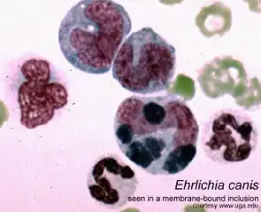
Ehrlichia canis seen in a membrane-bound inclusion (morulae) within the cytoplasm of a monocyte (buffy coat smear, Wright stain).
Samples:
Notes: Send all samples at room temperature, or preserved in sample buffer.
Interpretation of PCR Results:
Ehrlichia canis/ewingii
Ehrlichiae are a group of small, gram-negative, pleiomorphic, obligate intracellular cocci that infect monocytes in various animal species and in humans. Canine ehrlichiosis (also known as canine rickettsiosis, canine hemorrhagic fever and canine typhus) is usually caused by Ehrlichia canis and E. ewingii, or less commonly by E. chaffeensis or E. equi (now Anaplasma phagocytophilum). German shepherd dogs are thought to be particularly affected by the disease, but cats and humans can also be infected.
Clinical Signs
Dogs contract ehrlichiosis from bites of the brown dog tick (Rhipicephalus sanguineous) or by blood transfusion from another infected dog. There are three stages of ehrlichiosis, each varying in severity. The acute stage, occurring several weeks after infection and lasting for up to a month, can lead to fever and lowered peripheral blood cell counts due to bone marrow suppression. The second stage, called the subclinical phase, has no outward signs and can last for the remainder of the dog’s life, during which the dog remains infected with the organism. Some dogs eliminate the disease during this time, whereas others progress to the third and most serious stage of infection, the chronic phase. Very low blood cell counts (pancytopenia), bleeding, bacterial infection, lameness, neurological and ophthalmic disorders, and kidney disease, can result. Chronic ehrlichiosis can be fatal (Skotarczak, 2003).
Standard Diagnostic Methods
Ehrlichiosis is diagnosed most commonly by serologic testing of the blood for the presence of antibodies against the Ehrlichia organisms. However, during the acute phase of infection, the test can be falsely negative because the body has not yet had time to produce antibodies. Therefore, repeated testing is required. Ehrlichiosis can also be diagnosed by microscopic examination of a blood smear for the presence of the Ehrlichia morulae, which sometimes can be seen as intracytoplasmic inclusion bodies within a white blood cell. In recent years qualitative PCR has been used to detect presence of Ehrlichia (Gal et al., 2007).
Our Method
The Molecular Diagnostics Laboratory at Auburn University has developed a quantitative PCR approach targeting the 16S rRNA gene of all Ehrlichia spp. that detects ehrlichiosis with higher sensitivity than any other test (as few as 7 organisms per ml blood). E. canis, E. ewingii, and E. chaffeensis are differentiated by analysis of the product melting curves after PCR amplification.