
E. coli O157:H7
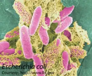
Scanning electron micrograph of E. coli bacteria on the surface of cultured cells.
Samples:
Notes: Send all samples at room temperature, preferably preserved in sample buffer.
Interpretation of PCR Results:
Eschericia (E.) coli
Escherichia (E.) coli is a gram-negative, facultatively anaerobic, straight bacterium. It is an important component of the biosphere, and serves a useful function in the body by suppressing the growth of harmful bacterial species and by synthesizing appreciable amounts of vitamins. E. coli colonizes the lower gut of animals and survives when released to the natural environment, allowing widespread dissemination to new hosts. Pathogenic E. coli strains are responsible for infection of the enteric, urinary, pulmonary and nervous systems (Dodd et al., 2003). E. coli O157:H7 is a foodborne pathogen, and is a verotoxin–producing serotype of E. coli, and is responsible for many cases of hemorrhagic colitis. E. coli O157:H7 infections occur worldwide.
Clinical Signs
E. coli O157:H7 bacteria is believed to mostly reside in the intestines of cattle but has also been found in the intestines of chickens, deer, sheep, and pigs (Dodd et al., 2003). The animals are merely the reservoir for the bacteria, and E. coli O157:H7 does not cause disease in carrier animals. However, foodborne transmission of E. coli O157:H7 from asymptomatic carrier animals to humans may cause severe human disease. This disease is hemorrhagic colitis characterized by cramps, abdominal pain, and watery diarrhea followed by bloody diarrhea. A low-grade fever may be present or absent in the initial stages. Dehydration is possible. Most infections are usually self-limiting and last about a week, and in a small percentage of cases serious, occasionally lethal complications are seen such as hemolytic uremic syndrome (HUS) and thrombotic thrombocytopenic purpura (TTP).
Standard Diagnostic Methods
E. coli O157:H7 infections are usually diagnosed by isolating the organism from fecal samples or testing the feces for E. coli verotoxin. E. coli O157:H7 is not detected in routine cultures but can be recognized by incubation on sorbitol– MacConkey agaror the variant cefeximine potassium telluride sorbitol-MacConkey agar. Hemorrhagic colitis agar is used to isolate bacteria from food samples. However, like all cultures, diagnosis is slow using this method, and more rapid diagnosis is possible using PCR techniques. Newer technologies using fluorescent and antibody detection are also under development.
Our Method
The Molecular Diagnostics Laboratory at Auburn University has developed a quantitative PCR technology targeting the eaegene, avirulence marker for the attachment and effacement present only in E. coli O157:H7 (Sheng et al., 2006). The PCR is designed and capable of detecting a single copy of this target gene in the PCR nucleic acid input.