
Hemotrophic Mycoplasmosis (canine/feline)
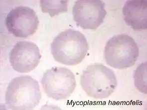
Mycoplasma haemofelis in a blood film from an infected cat. (Wright Stain, ×1,200)
Samples:
Notes: Send all samples at room temperature, or preserved in sample buffer.
Interpretation of PCR Results:
Mycoplasma haemofelis/haemocanis
Mycoplasmas are the smallest free-living microorganisms. They are currently divided into hemotrophic and nonhemotrophic types. Hemotrophic mycoplasmas are gram-negative, nonacid-fast, epicellular parasitic bacteria of erythrocytes. Mycoplasma haemofelis (formerly known as Haemobartonella felis) and Mycoplasma haemocanis (formerly known as Haemobartonella canis) can attach to the surface of erythrocytes of cats and dogs, respectively, and cause hemolytic anemia through extravascular destruction of erythrocytes by the mononuclear phagocyte system and intravascular lysis (Kewish et al., 2004).
Clinical Signs
The severity of disease produced by M. haemofelis varies, with some cats having mild anemia and no clinical signs and some having marked depression and severe anemia leading to death. Most common clinical signs are tachypnea, depression, weakness, anorexia, weight loss, pale mucous membranes, dehydration, icterus, and splenomegaly. In dogs, however, most non-splenectomized dogs infected with M. haemocanis do not develop clinical evidence of disease. Splenectomized dogs infected become listless and develop pale mucous membranes as the anemia progresses but generally have normal rectal temperatures and appetites.
Standard Diagnostic Methods
The only possible diagnostic procedure before the arrival of molecular diagnosis was microscopic examination of blood smears. This procedure has many drawbacks, however, since bacterial pathogens may be confused with artifacts or lost after EDTA treatment of collected blood (Criado-Fornelio et al., 2003). Many PCR-based methods have been developed to detect presence of M. haemofelis or M. haemocanis that provided remarkable sensitivity and specificity.
Our Method
The Molecular Diagnostics Laboratory at Auburn University has developed a quantitative PCR approach targeting the 16S rRNA gene of Mycoplasma spp. that detects both M. haemofelis and M. haemocanis with high sensitivity (as low as 7 genome copies per ml of blood) and specificity.