
Pseudomonas aeruginosa
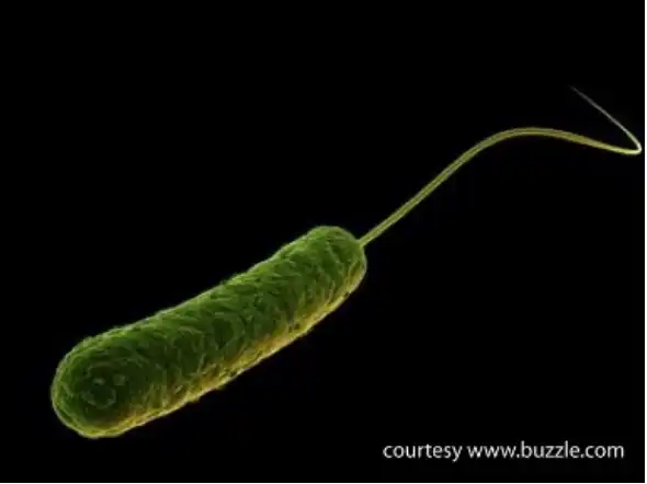
Artistic rendering of a Pseudomonas aeruginosa bacterium.
Samples:
Notes: Send all samples at room temperature, preferably preserved in sample buffer.
Interpretation of PCR Results:
Pseudomonas (P.) aeruginosa
Pseudomonas (P.) aeruginosa is a gram-negative, aerobic, rod-shaped bacterium. It is ubiquitous in soil and water but occurs regularly on the surfaces of plants and occasionally on the surfaces of animals. Pseudomonas aeruginosa is metabolically extremely versatile, forms highly resistant biofilms on moist surfaces, and has become increasingly recognized as an emerging opportunistic pathogen of clinical relevance. Several epidemiological studies track its occurrence as a nosocomial pathogen and indicate that antibiotic resistance is rapidly increasing in clinical isolates. P. aeruginosa is also recognized as serious contaminant of pharmaceutical products.
Clinical Signs
P. aeruginosa infections typically are contracted from heavily infested surfaces such as tubing or water tubs, and virtually any animal can contract these opportunistic infections. Depending on the site of infection, clinical symptoms typically appear as localized purulent inflammation. Secondarily, these infections may become disseminated via the bloodstream, leading to symptoms of severe or fatal gram-negative septicemia such as disseminated intravascular coagulation. Cystic fibrosis patients frequently suffer from pneumonia caused by P. aeruginosa. Similarly, patients with burn wounds or immunocompromised individuals frequently contract localized or systemic P. aeruginosa infections.
Standard Diagnostic Methods
P. aeruginosa infections are usually diagnosed by isolating the organism from clinical or environmental samples by standard bacteriological culture on blood agar plates. However, like all cultures, diagnosis is slow using this method, and more rapid diagnosis is possible using PCR techniques. Alternative technologies using fluorescent and antibody detection have also been developed, but are not highly sensitive.
Our Method
The Molecular Diagnostics Laboratory at Auburn University has developed a quantitative PCR technology targeting the oprL gene, a peptidoglycan-associated lipoprotein present only in pathogenic Pseudomonas species (Deschaght et al., 2009). The PCR is designed and capable of detecting a single copy of this target gene in the PCR nucleic acid input, and differentiating the P. aeruginosa amplicon from all other Pseudomonas species.