
Researchers Using Innovative Blood Testing in Breast Cancer Detection
Much of medical science derived for human treatment programs is based on earlier research involving animal models. A team of scientists at the College of Veterinary Medicine has received a grant to support studies applying human medical knowledge to dogs in the detection of breast cancer.
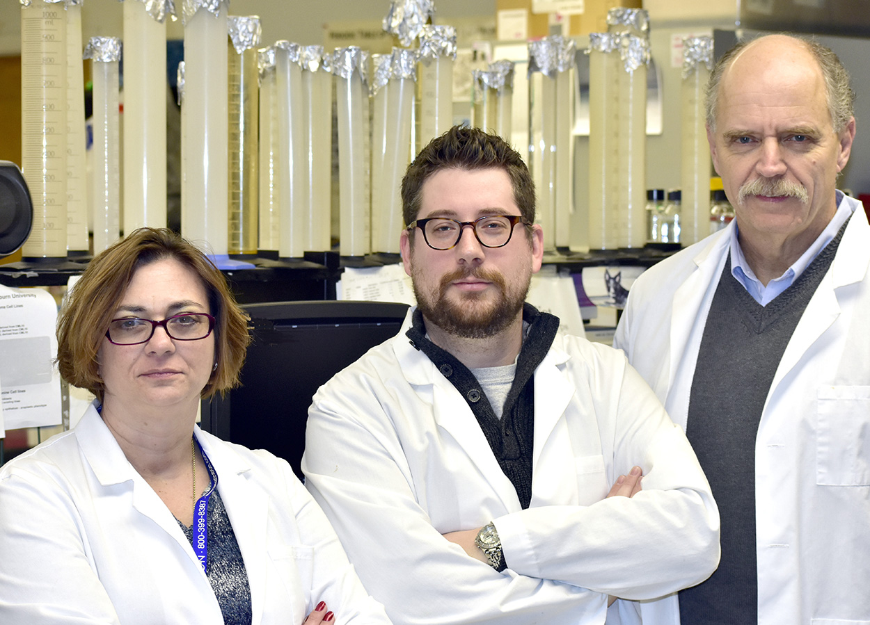
Dr. Eric Fish, a clinical lecturer and doctoral candidate in the Department of Pathobiology, in collaboration with Dr. Annette Smith, the Lowder Distinguished Professor in the Department of Clinical Sciences, and Dr. Curt Bird, a professor in the Department of Pathobiology, recently received a $28,458 grant from the American College of Veterinary Internal Medicine (ACVIM) Foundation for their study titled, “Circulating MicroRNA as Predictive Biomarkers for Canine Mammary Neoplasia.” Their research explores if microRNA blood markers can be used to accurately detect breast cancer in dogs and also to predict how well patients will respond to treatment.
Breast cancer is common among dogs — just as in humans — and the disease shares characteristics among both at the genetic level, according to Dr. Fish.
“MicroRNA as a marker has been in use in human medicine for some five or so years,” Dr. Fish said. “But it really has not been used clinically with veterinary medicine.”
The CVM team is building on the human research model to apply the science to dogs.
The dog parallels well with the human breast cancer model, according to the CVM team. Although it is less common among spayed animals – particularly dogs spayed at an early age, which rarely develop breast cancer – “intact” dogs suffer breast cancer at much the same rate as do humans.
Dogs possess a number of other characteristics that make the dog a good model for this research, according to the CVM researchers.
“The genetics of the immune system are structured similarly (as in humans),” Dr. Fish says. “Dogs have a similar course in the breast cancer disease distribution of severity as do humans. Their larger size (as compared to smaller research animals) makes the dog model a better representation for drug and blood/tissue sampling, and such characteristics as hormonal conditions, age and obesity work in a dog’s system much as they work in a human.”
Screening involves a routine blood test. The blood is examined for the microRNA markers. And according to the research team, the simplicity of the procedure is the uniqueness of their work. The researchers explain their project:
“Normal breast tissue and less aggressive types of breast cancer rely on the hormones estrogen and progesterone for growth and development. In human medicine, anti-hormone compounds can be used to treat low-grade breast cancer, and in dogs, surgical removal of the ovaries and/or uterus can reduce the effects of these hormones. However, as these tumors become more cancerous and less differentiated, they can begin to grow independently of these normal signals. Therefore, loss of estrogen and progesterone receptors can be seen as a proxy marker of a more dangerous type of breast cancer. At the same time, over-expression of a receptor called HER-2 can push these tumors into overdrive and make them very hard to treat.”
“The exciting thing about our research is that early results suggest a number of these microRNA correlate with—and may actually drive changes in—the hormone receptor expression profile, and thus the biological behavior of the tumor,” Dr. Fish said. “Those markers may help tell us non-invasively if breast cancer is present. We also are studying this to see if those markers also can tell us more about the specific type of cancer, how aggressive the type is, and how it will respond to specific treatments. We believe that if this research is successful, it will provide an early detection method that is minimally invasive for the patient.”
The project has been underway for about 18 months, according to Dr. Fish. The team currently is working on its first peer-reviewed paper, which is on track to be published in early 2017.
“The goal of the research is to improve the overall survival of breast cancer in dogs through earlier and minimally invasive detection of cancer, tumor recurrence and metastasis,” Dr. Fish said.
“We are very pleased for the opportunity to perform research in the exciting area of microRNAs,” adds Dr. Smith, who was instrumental in obtaining the research grant. “These small molecules may provide clues to early diagnosis and prognosis in canine mammary tumors with just a small blood sample.
“Ultimately, therapeutics blocking some of these molecules may also be developed. There will likely be transitional applications for human breast cancer medicine. We appreciate the ACVIM Foundation’s funding of this project.”
The ACVIM Foundation exists to bridge the gap between available funding and the vital work that needs to be done. Because clinical studies in veterinary medicine are severely underfunded and receive virtually no government support, ACVIM specialists have long depended on the generosity of private donors and industry sponsors to support their research.
“The Auburn University study coincides with the mission of the ACVIM Foundation, which is to improve animal and human health by funding discovery and education,” said Andrea Miller, foundation director. “Dr. Annette Smith and her collaborators are leaders in veterinary medicine, and we are honored to support their valuable research.”
Animal Healthcare Bailey Small Animal Teaching Hospital Research