
Leptospira spp.
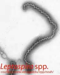
Scanning electron micrograph of Leptospira spp. Notice the corkscrew appearance of the
bacterium.
Samples:
Notes: Send all samples at room temperature, or preserved in sample buffer (PDF)
Interpretation of PCR Results:
Leptospira
Leptospirosis is a zoonotic disease of worldwide veterinary significance in many animal species. It is caused by infection with antigenically distinct serovars of the spirochete bacterium Leptospira interrogans sensu lato, of which eight are of greatest importance to dogs and cats (Levett, 2001). There are multiple, antigenically distinct organisms within each serogroup, which are referred to as serovars. Serovars are maintained in nature in numerous subclinically infected wild and domestic animal reservoir hosts that serve as potential sources of infection and illness for humans and other incidental hosts.
Clinical Signs
Leptospira organisms are passed in urine and penetrate mucous membranes or abraded skin and multiply rapidly upon entering the blood vascular space. The bacteria continuously spread in the body and replicate further in many tissues including kidney, liver, spleen, central nervous system (CNS), eyes, and genital tract. Thereafter, increased serum antibodies clear the spirochetes from most organs, but bacteria may persist in the kidneys and be shed in urine for weeks to months. The extent of damage to internal organs is variable depending on the virulence of the organism and host susceptibility. Chronic active hepatitis has been a consequence of L. interrogans serovar grypotyphosa infection in dogs.
Standard Diagnostic Methods
Diagnosis of leptospirosis is based on a combination of suggestive historical information, physical findings, nonspecific laboratory findings, and confirmatory testing. Confirmatory tests include serologic testing to detect antibody production to Leptospira spp. Very high antibody titers are suggestive of infection, but paired serum titers produce more reliable prognostic information. Direct detection of the bacterium may be done by culture of urine or blood culture, identification of leptospiral DNA, FA staining of urine, or urine dark-field microscopy. Culture of Leptospira spp. can be difficult and time-consuming.
Our Method
The quantitative PCR approach we have developed uses the highly conserved ligA and ligB genes as the amplification target (Raghavan et al, 2005), and detects, by fluorescent probes, single copies of conserved sequences of the genes present in the sample input to the PCR. In a study by Xu et al. (Xu et al., 2014), our pathogenic Leptospira spp. PCR has been proven the most sensitive detection method for Leptospira infection in dogs.